Assisted Reproductive Technology Support System for Infertility Treatment(FiTTE:Fertility image Testing Through Embryo)
FiTTE features two functions (systems) related to infertility treatment that directly address challenges in clinical practice.
Overview of Fertility Treatment
Despite a declining number of live births in Japan, the number of live births resulting from assisted reproductive technology (ART)—including in vitro fertilization, intracytoplasmic sperm injection, egg and embryo cryopreservation, fresh embryo transfer, and frozen embryo transfer—has increased due to technological advances and later marriage. As of 2018, one in every 16 newborns was conceived through ART (See figure below).
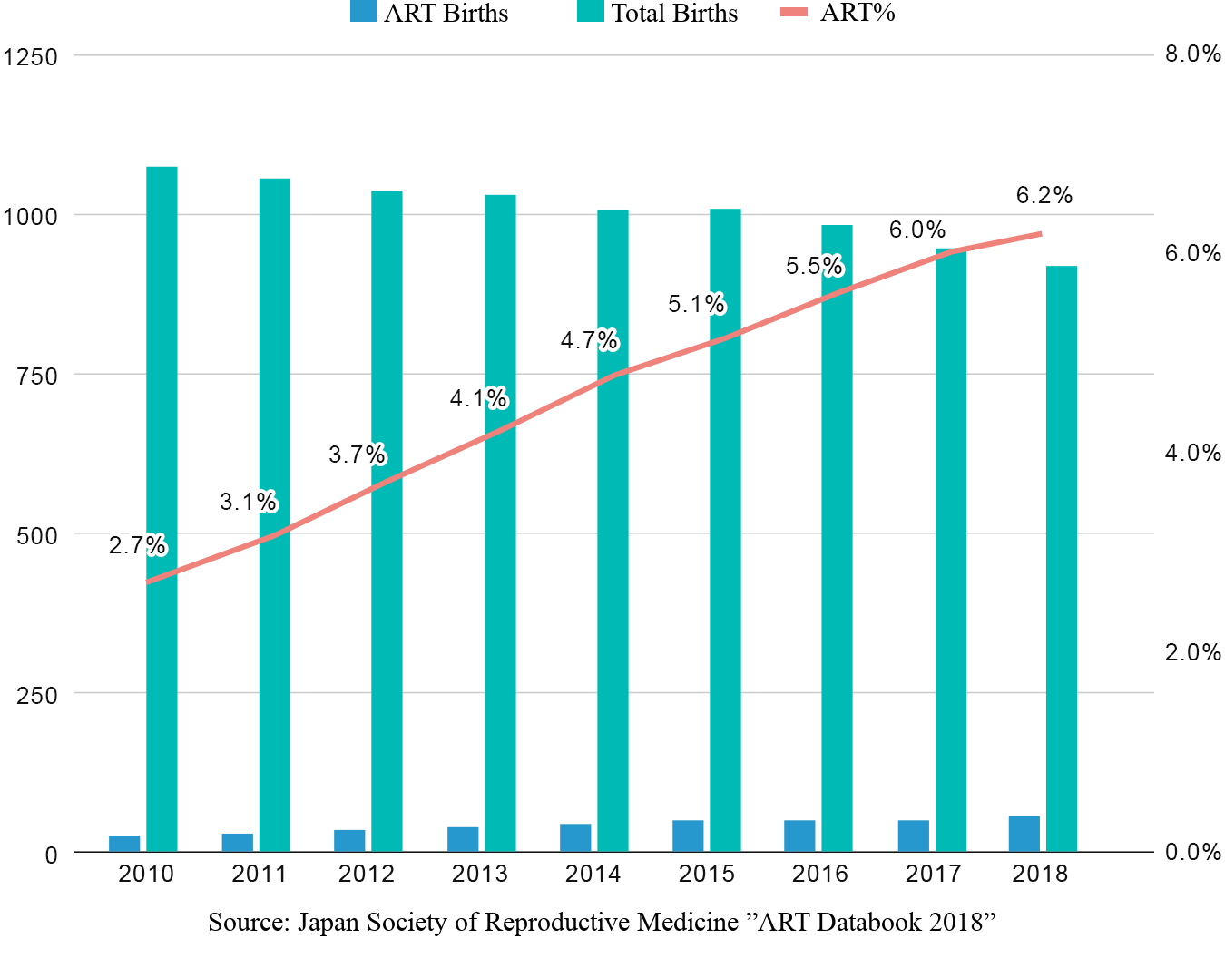
In Japan, where infertility treatment technology has advanced, such treatments are becoming increasingly common, with one in every 5.5 couples reportedly undergoing them.
Although insurance coverage for infertility treatments was expanded in April 2022, the significant financial burden on patients remains a major issue, partly due to certain restrictions.
[ Function 1] Non-Invasive Determination System of Chromosome Aneuploidy in Embryos by Time-Lapse Image Analysis
Clinical Challenge
Embryo Aneuploidy,known cause of recurrent miscarriage, is typically tested using
PGT-A (Preimplantation Genetic Testing for Aneuploidy).
This technique requires invasive procedures on the embryo and raises concerns about its impact on pregnancy and live birth rates. Consequently, its active use in fertility clinics has been the subject of extensive debate, including discussions within the Japan Society of Obstetrics and Gynecology.
The implementation cost also tends to be high, resulting in a significant burden on patients.
Our Technology
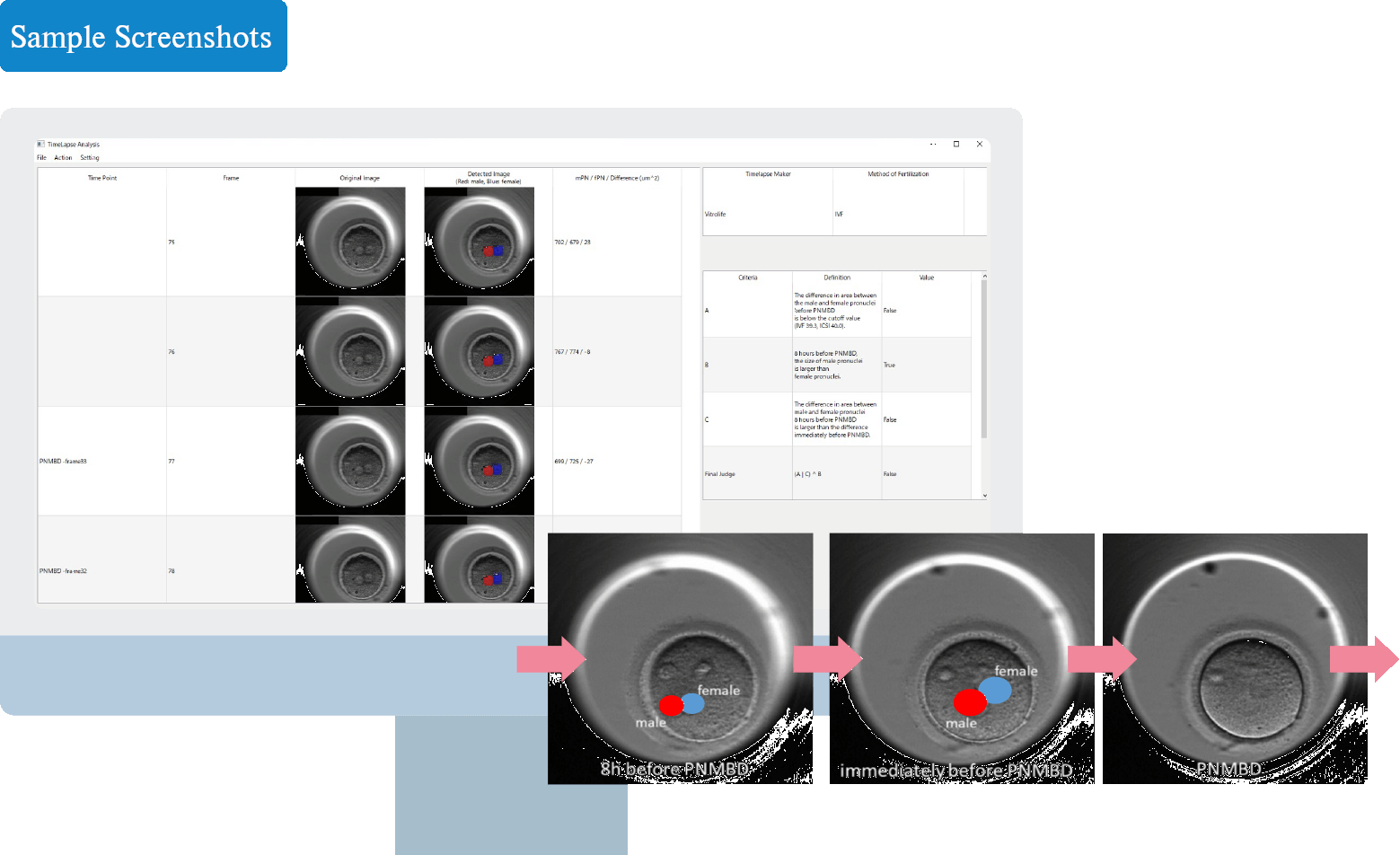
We have developed a system that automates the non-invasive detection of chromosomal aneuploidy based on embryonic developmental characteristics (time point identification, area calculation) using our proprietary image segmentation AI technology. This method was originally developed in collaboration with partner clinics and academic institutions (Ootsuki et al. Fertility and Sterility. 2019)
A retrospective observational study was conducted on approximately 100 cases and tens of thousands of images. Using time-lapse videos labeled by embryologists as training data, an AI model for image segmentation was developed, enabling the extraction of embryonic development process features (time point identification, area calculation).
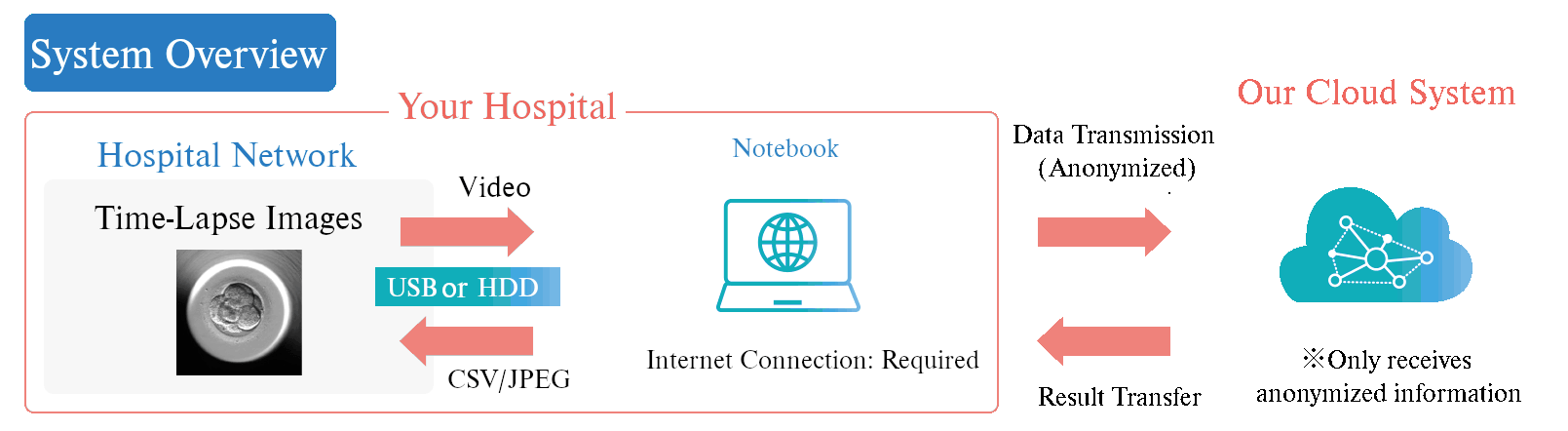
By loading time-lapse images of in vitro fertilization and ICSI embryo culture onto software connected to an image segmentation AI hosted on a cloud server, we provide physicians with chromosomal aneuploidy detection information.
This enables inexpensive, non-invasive, and rapid testing within the clinic to provide necessary information. (※ Physicians will also review other clinical information and make a comprehensive judgment.)

What is Chromosome Aneuploidy Detection Software?
We are developing aneuploidy detection software in the field of assisted reproductive
technology for infertility treatment. While conventional preimplantation genetic testing for
aneuploidy (PGT-A) is a method for detecting chromosomal abnormalities believed to cause
recurrent miscarriage, our product offers the following advantages over conventional methods:
- Non-invasive (potential for improved pregnancy rates)
- Immediate results
- Low-cost implementation
- In a retrospective study, although based on a small dataset, it has already demonstrated accuracy comparable to PGT-A (※)
(Presented at the 66th Annual Meeting of the Japan Society for Reproductive Medicine in 2021)
※What is PGT-A?
PGT-A is a test that examines the chromosome count of embryos obtained through in vitro fertilization before implantation.
It is an effective screening method to prevent miscarriages associated with age-related factors.
While this test is expected to reduce the risk of miscarriage and improve pregnancy rates,
important considerations include the fact that the test's accuracy is not 100%, and that a normal embryo may not necessarily be found after testing.

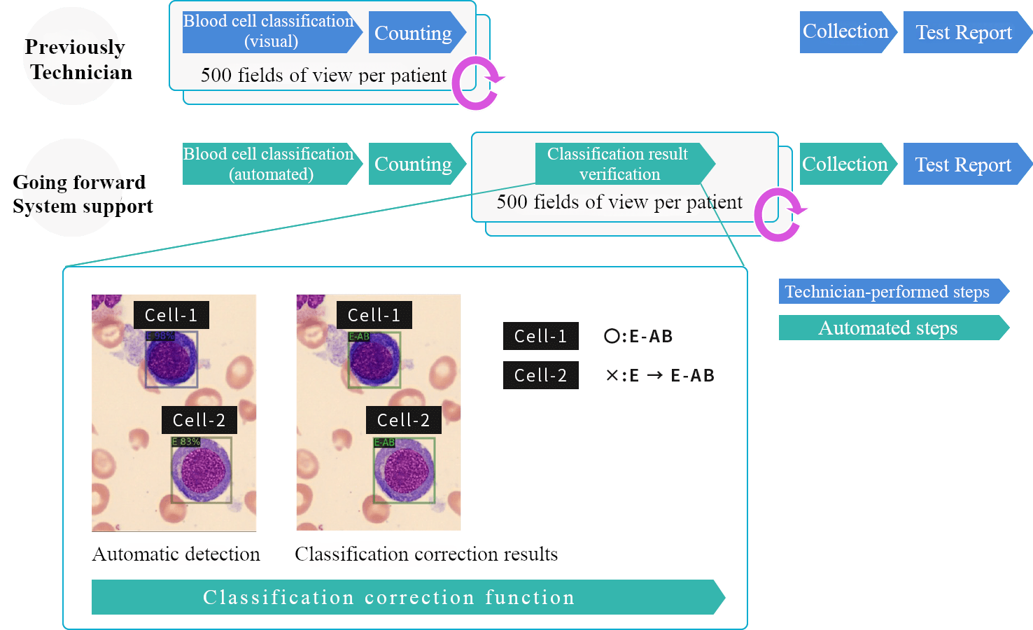
Function Details
- Collected approximately 100 cases and tens of thousands of images through retrospective observational studies (ongoing)
- Trained on time-lapse images labeled by embryologists and physicians
- Performed dynamic analysis of male and female pronuclei (identifying disappearance points, calculating area)
- Implemented a system for pseudonymizing and processing data within the hospital
[ Function2 ] Predicting system for pregnancy and live birth using time-lapse embryo imaging analysis
Clinical challenges
In assisted reproductive technology (ART), including in vitro fertilization and intracytoplasmic sperm injection,the condition of the embryo is considered crucial for subsequent pregnancy and live birth.
The current primary method involves visually assessing embryo morphology at the time of transfer. While various evaluation techniques, such as analyzing the embryo's developmental process, are being discussed in relevant academic societies, no established method for predicting pregnancy and live birth has yet been developed.
Our Technology
We developed an image analysis AI that outputs a five-level assessment of pregnancy and live birth potential by inputting time-lapse images of the embryo culture process.
As a retrospective observational study involving approximately 20,000 cases, we constructed the image analysis AI by extracting embryonic development processes and morphological characteristics from the correlation between time-lapse images of the embryo culture process and pregnancy/live birth outcomes.
By loading time-lapse images of embryos cultured via IVF or ICSI into software connected to the cloud-based image analysis AI, it provides physicians with a 5-point grading result, supporting the selection of embryos for transfer.
This software facilitates physician-assisted embryo transfer (physicians also review other clinical information for comprehensive decision-making). The image analysis AI model is deployed on the cloud and integrated with this software.